Home
Uncategories
Rib Cage Muscles Diagram / External Intercostals Anatomy Function And Treatment - It is flexible and can expand and contract by the action of the muscles of respiration.
Rib Cage Muscles Diagram / External Intercostals Anatomy Function And Treatment - It is flexible and can expand and contract by the action of the muscles of respiration.
Rib Cage Muscles Diagram / External Intercostals Anatomy Function And Treatment - It is flexible and can expand and contract by the action of the muscles of respiration.. Each are symmetrically paired on a right and left side. These muscles connect the lower part of the spine to the ilium and the femur and. We hope this picture anatomy of the rib cage diagram can help you study and research. Anatomynote.com found anatomy of the rib cage diagram from plenty of anatomical pictures on the internet. The human rib cage is made up of 12 paired rib bones;
Muscles that helpful in expanding the thoracic cavity are called the inspiratory muscles. Womens body parts stomach 4 photos of the womens body parts stomach body diagram stomach, body parts digestive system, body parts in stomach area, body parts liver, body parts spleen, human body parts stomach, woman body organs, woman body parts found, stomach, body diagram stomach, body parts digestive system, body. In most of the less severe cases, the pain around rib cage results from strained muscles due to coughing, overstretching or postural changes. Related posts of rib cage diagram with organs womens body parts stomach. These muscles attach the upper limb to the axial skeleton of the trunk and support the.
Anatomy Of The Rib Cage Diagram from www.anatomynote.com These muscles attach the upper limb to the axial skeleton of the trunk and support the. The muscles of the thoracic cage are the pectoralis major, pectoralis minor, serratus anterior, subclavius, intercostal (external, internal and innermost), subcostal and transversus thoracis muscles, including the diaphragm. Each pair is numbered based on their attachment to the sternum, a bony process at the front of the rib cage which serves as an anchor point. A group of muscles connected to the rib cage, which help stabilize the shoulder. We think this is the most useful anatomy picture that you need. The human rib cage is a component of the human respiratory system. The intercostal muscles have different layers that are attached to the ribs to help build the chest wall and. Anatomynote.com found anatomy of the rib cage diagram from plenty of anatomical pictures on the internet.
There are 11 pairs of external intercostal muscles.
Originate at the lower border of the rib, inserting into the superior border of the rib below. Stretching out muscles under the rib. Introduction edit . Anatomy of the upper back and middle back (thoracic spine) your upper and middle back starts from just below your neck and extends to about 5 inches below your shoulder blades. Muscles of the thoracic cage. The human rib cage is a component of the human respiratory system. The thoracic cage (rib cage) is the skeleton of the thoracic wall. It provides a strong framework onto which the muscles of the shoulder girdle, chest, upper abdomen and back can attach. Muscles labeled front and back 12 photos of the muscles labeled front and back muscle diagram labeled front and back, muscle system labelling (front and back), muscular. The first rib is the widest, shortest and has the sharpest curve of all the ribs. It is formed by the 12 thoracic vertebrae, 12 pairs of ribs and associated costal cartilages and the sternum. It can also be a sign of. The thoracic cage takes the form of a domed bird cage with the horizontal bars formed by ribs and costal cartilages.
The human rib cage is a component of the human respiratory system. When you go to sleep, position 2 to 3 pillows under your head and 1 under your upper back to slightly elevate your torso. Stretching out muscles under the rib. With the upper ribs, closer to the nodule (and in the case of lower ribs, a little further from the nodule) they are curved and have a rough surface that connects them with muscles, angulus costae. There are various causes for pain around rib cage and the presentation of symptoms also varies according to the cause.
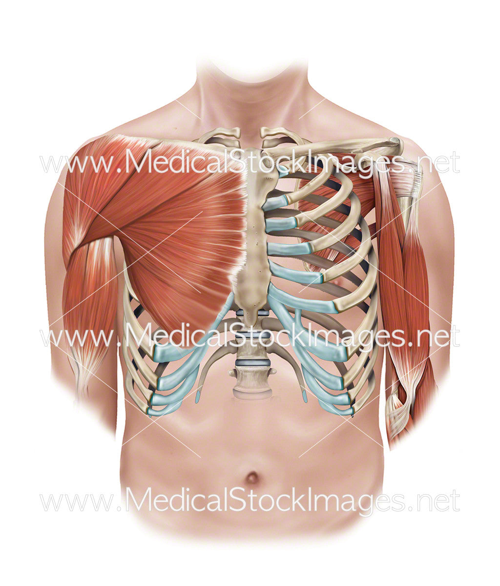
Superficial And Deep Muscles Of The Shoulder And Rib Cage Medical Stock Images Company from cdn.shopify.com The head only articulates with the body of the t1 vertebra and therefore only one articulatory surface is present. With the upper ribs, closer to the nodule (and in the case of lower ribs, a little further from the nodule) they are curved and have a rough surface that connects them with muscles, angulus costae. Knowing what can affect your rib cage, back muscles, and ligaments that support the spine can help to take steps to relieve the pain. This muscle lies between the back of your rib cage and your shoulder blade near the subscapularis (one of the four rotator cuff muscles). Each are symmetrically paired on a right and left side. It encloses the thoracic cavity, which contains the lungs. There are various causes for pain around rib cage and the presentation of symptoms also varies according to the cause. Intercostal muscles support respiratory function, while the upper abdominal muscles support your.
Intercostal muscle strain is an injury affecting the muscles between two or more ribs.
Muscles labeled front and back 12 photos of the muscles labeled front and back muscle diagram labeled front and back, muscle system labelling (front and back), muscular. Of all 24 ribs, the first seven pairs are often labeled as 'true.' The muscles on the left side are the superficial muscles (close to the surface), and the muscles on the right are positioned beneath the superficial muscles. It encloses and protects the heart and lungs. Originate at the lower border of the rib, inserting into the superior border of the rib below. There are 11 pairs of external intercostal muscles. These muscles attach the upper limb to the axial skeleton of the trunk and support the. Human anatomy for muscle, reproductive, and skeleton. The human rib cage is a component of the human respiratory system. The area under the ribs consists of intercostal muscle, ligaments and tendons, as well as the abdominal obliques, transverus abdominis and rectus abdominis just below the rib cage. The rib cage is a bony structure found in the chest (thoracic cavity). Located in the rib cage, this muscle keeps the shoulder blade against the chest wall and helps rotate the shoulder blade upward. Intercostal muscles support respiratory function, while the upper abdominal muscles support your.
Muscles labeled front and back 12 photos of the muscles labeled front and back muscle diagram labeled front and back, muscle system labelling (front and back), muscular. Rib cage anatomy the rib cage, shaped in a mild cone shape and more flexible than most bone sets, is made up of varying elements such as the thoracic vertebra, 12 equally paired ribs, costal cartilage, and held together anteriorly by the sternum. An inhalation is accomplished when the muscular diaphragm, at the floor of the thoracic cavity, contracts and flattens, while the contraction of intercostal muscles lift the rib cage up and out. As in the typical ribs, the tubercle has a facet for articulation with the transverse process of vertebrae. It is formed by the 12 thoracic vertebrae, 12 pairs of ribs and associated costal cartilages and the sternum.
Male Diaphragm Muscle With Circulatory System In Rib Cage Stock Photo Download Image Now Istock from media.istockphoto.com In most of the less severe cases, the pain around rib cage results from strained muscles due to coughing, overstretching or postural changes. It is flexible and can expand and contract by the action of the muscles of respiration. Stretching out muscles under the rib. Rib cage, in vertebrate anatomy, basketlike skeletal structure that forms the chest, or thorax, and is made up of the ribs and their corresponding attachments to the sternum (breastbone) and the vertebral column.the rib cage surrounds the lungs and the heart, serving as an important means of bony protection for these vital organs.in total, the rib cage consists of the 12 thoracic vertebrae and. As in the typical ribs, the tubercle has a facet for articulation with the transverse process of vertebrae. All muscles that are attached to the human rib cage have the inherent potential to cause a breathing action. Each are symmetrically paired on a right and left side. The rib cage has three important functions:
The intercostal muscles have different layers that are attached to the ribs to help build the chest wall and.
We think this is the most useful anatomy picture that you need. It can also be a sign of. Several muscles that move the arms, head, and neck have their origins on the sternum. Muscles are groups of cells in the body that have the ability to contract and relax. In most of the less severe cases, the pain around rib cage results from strained muscles due to coughing, overstretching or postural changes. Muscles that helpful in expanding the thoracic cavity are called the inspiratory muscles. Related posts of rib cage diagram with organs womens body parts stomach. These muscles attach the upper limb to the axial skeleton of the trunk and support the. This muscle lies between the back of your rib cage and your shoulder blade near the subscapularis (one of the four rotator cuff muscles). With the upper ribs, closer to the nodule (and in the case of lower ribs, a little further from the nodule) they are curved and have a rough surface that connects them with muscles, angulus costae. Knowing what can affect your rib cage, back muscles, and ligaments that support the spine can help to take steps to relieve the pain. The sternum, commonly known as the breastbone, is a long, narrow flat bone that serves as the keystone of the rib cage and stabilizes the thoracic skeleton. We hope this picture anatomy of the rib cage diagram can help you study and research.
Muscles are groups of cells in the body that have the ability to contract and relax rib cage muscles. These muscles attach the upper limb to the axial skeleton of the trunk and support the.


0 Comments:
Posting Komentar